Foramen Lacerum
Definition
Foramen lacerum is a hole that is found at the base of the skull, with a characteristic triangular shape. These are the elements that surround the foramen lacerum and practically contribute to its formation: sphenoid bone (anterior border), petrous temporal bone (more exactly, its apex) and the occipital bone (specifically, the basilar part). It is known that foramen lacerum is located anteriorly and medially from the carotid canal. The hole is covered by cartilage (connective tissue) in the postnatal period. In general, the foramen lacerum has a length of 9 mm and a breadth of 7 mm. The foramen lacerum is found on each side of the skull base, at a close distance from the pharyngeal tubercle of the occipital bone.
Function of Foramen Lacerum
The internal carotid artery appears at a superior point from the foramen lacerum, after having passed from the carotid canal into the base of the skull. Even though it exists the carotid canal, the internal carotid artery is not going to pass through the foramen lacerum. However, because it is located at a close distance, the part that is found above the foramen lacerum is also known as the lacerum segment. These are the elements that pass through the foramen lacerum: venous drainage, nerve and artery of the pterygoid canal.
The foramen lacerum is also transited by the greater petrosal nerve. This will eventually become a part of the nerve of the pterygoid canal (this also contains the deep petrosal nerve). This nerve contains both sympathetic and parasympathetic fibers, covering the innervation for the blood vessels in the area, as well as for the mucous membranes, salivary and lacrimal glands.
A terminal branch of the ascending pharyngeal artery also passes through the foramen lacerum, as well as emissary veins. The latter are responsible for connecting the intracranial cavernous sinus with the extracranial pterygoid plexus; unfortunately, due to this connection, there is also a high risk for an infection traveling from one point to the other. Also, the foramen lacerum offers the invasive nasopharyngeal carcinoma with the perfect access point into the cavernous sinus. It is because of this connection that the cranial nerves are affected in the spread of the nasopharyngeal carcinoma.
Studies performed on cadavers have demonstrated that the foramen lacerum is not a true foramen. Scientists have based this affirmation on the fact that no significant structures go above the fibrous tissue that covers it. It was suggested that this is rather an extension of the lacerum portion of the carotid canal and not a foramen per say.
In conclusion, the structures that pass at the whole length of the foramen lacerum are the meningeal branch of the ascending pharyngeal artery and the emissary veins. The other structures that traverse the foramen lacerum only partially include the internal carotid artery and the greater petrosal nerve.
Pictures
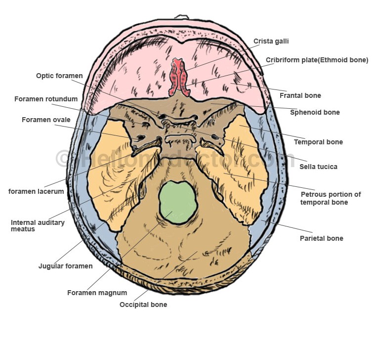
Foramen Lacerum Picture 1 : Floor of the cranial cavity showing various parts including the Foramen lacerum, Optic foramen, Foramen rotundum, Foramen ovale, Internal auditory meatus, Jugular foramen, Foramen magnum, Occipital bone, Parietal bone, Petrous portion of temporal bone, Sella turcica, Temporal bone, Sphenoid bone, Frontal bone, Cribriform plate (Ethmoid bone) and Crista galli.
Foramen lacerum syndrome
This condition is actually represented by the aneurysm of the internal carotid artery. A congenital condition, it generally involves the intradural portion of the respective artery. Among the symptoms that patients present, there are: inflammation of the meninges, orbital headache and migraines.
