Definition: What is Hilar adenopathy?
Hilar adenopathy can be defined as the enlargement of the lymph nodes, occurring at the level of the pulmonary hilum. This condition does not appear on its own, always signifying the existence of an underlying pathology. It is possible that both lungs present the enlargement of the lymph nodes.
Hilar Adenopathy Causes
These are the potential causes or etiologies that can lead to the appearance of the hilar adenopathy:
- Inflammatory
- Sarcoidosis
- Inorganic dust disease (silicosis, berylliosis)
- Malignant
- Lymphoma (more often Hodgkin lymphoma, in comparison to non-Hodgkin lymphoma)
- Metastases
- Bronchogenic carcinoma (primary hilar tumor)
- Mediastinal tumors
- Infectious
- Tuberculosis
- Histoplasmosis
- Infectious mononucleosis
- Fungal infection
- Mycoplasma
- Intestinal lipodystrophy (Whipple’s disease)
- Coccidioidomycosis
- Viral infection
- Atypical mycobacterial infection
- Tularemia
- Anthrax
- Extrinsic allergic alveolitis – bird fancier’s lung (hypersensitivity pneumonitis)
- Other causes (more rare)
- Churg-Straus syndrome
- HIV
- Still’s disease (adult onset)
- Reaction to medication.
The following conditions lead to the appearance of unilateral or bilateral, symmetrical hilar adenopathy: primary tuberculosis, fungal infection, atypical mycobacterial infection, viral infection, tularemia, anthrax, bronchogenic carcinoma, lymphoma, sarcoidosis and silicosis. On the other hand, the bilateral symmetrical adenopathy can only be caused by: sarcoidosis (most common), viral infections (adenovirus or mononucleosis).
Diagnosis
One of the most investigations used for the diagnosis of the hilar adenopathy is the plain pulmonary X-ray. However, has to take into consideration that the pulmonary arteries go through the same area. In the situation that these vessels are enlarged, they might be mistaken for hilar adenopathy. In order to make the difference between this condition and the enlarged vessels, you need to analyze their appearance. While the enlarged lymph nodes have a lumpy or bumpy appearance (characteristic opacity on the X-ray), the enlarged pulmonary arteries appear to be smooth (in reference to their contour).
Chest CT can also be used for the confirmation of hilar adenopathy. Depending on the results provided by the imaging studies, the enlargement of the lymph nodes can be classified as unilateral or bilateral. In the situation that the hilar adenopathy is bilateral, it can be further classified into symmetrical or asymmetrical.
Treatment
In order for the hilar adenopathy to disappear, the treatment has to be concentrated on the underlying etiology.
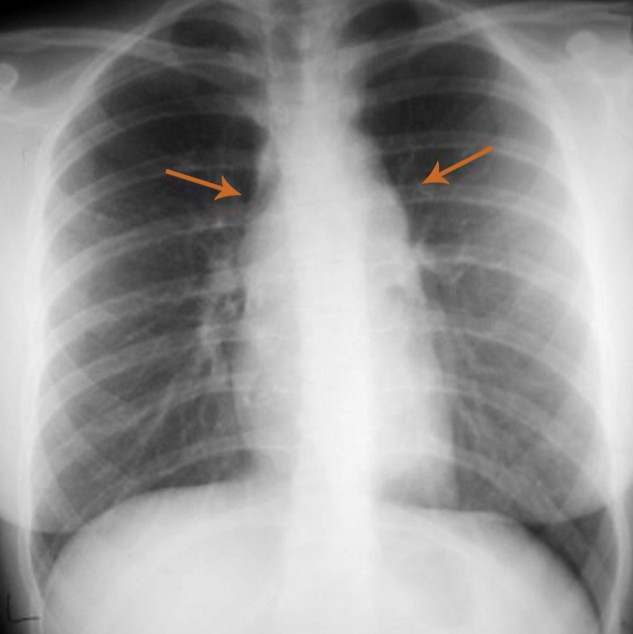
Pictures of Hilar Adenopathy
Take a look at how Hilar Adenopathy looks like: